Turning standard radiotherapy into a precision therapeutic weapon through nanoparticle activation.
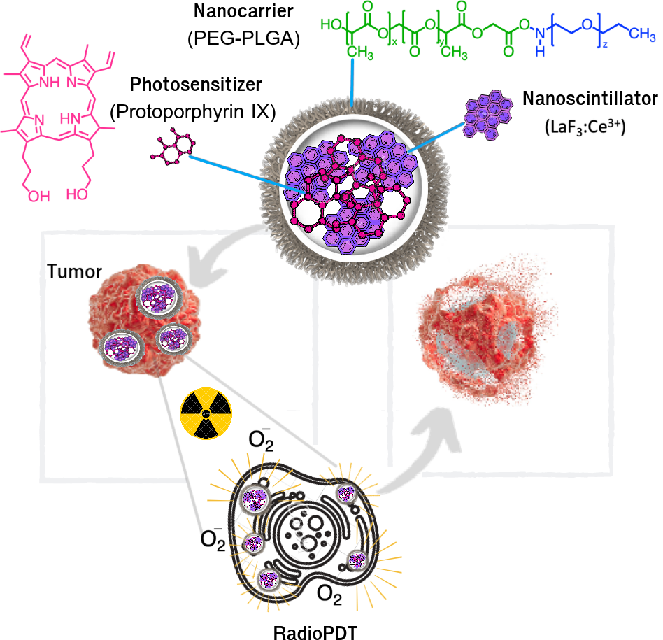
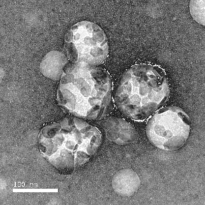
RadioLite's core technology is a novel class of radiation-activated nanoparticles designed to amplify the therapeutic effect of clinical radiotherapy while sparing healthy tissue. The platform is built on PEG-PLGA nanospheres (~100 nm diameter) that encapsulate both a scintillator core and a photosensitizer coating.
Unlike conventional photodynamic therapy (PDT), which requires visible light that cannot penetrate deep tissue, our nanoparticles are activated by ionizing radiation — the same radiation already used in cancer treatment. This means the therapy works deep inside the body, at the tumor site, without additional light sources or invasive procedures.
When ionizing radiation (X-rays, 6 MV) interacts with our proprietary LaF3:Ce3+ scintillator nanoparticles, it triggers a cascade of energy transfer events. The scintillator core absorbs X-ray energy and emits luminescence (UV/visible photons) within picoseconds. This luminescence is harvested via Förster Resonance Energy Transfer (FRET) to activate nearby Protoporphyrin IX (PPIX) photosensitizers.
The activated photosensitizers generate highly reactive oxygen species (ROS) — primarily singlet oxygen (1O2) and hydroxyl radicals (·OH). These ROS species are lethal to cancer cells. They damage DNA, disrupt cell membranes, and trigger apoptotic pathways. Because the ROS are generated locally at the nanoparticle surface (range ~40 nm), their effect is limited to the immediate tumor microenvironment, minimizing off-target effects.
Uniform particles, and the same chemistry from a bench batch to a production batch.
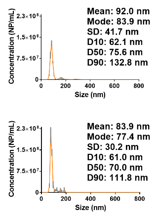
The particles come out the same size, batch after batch. Mean diameter ~92 nm with tight size distribution. Demonstrated >90% encapsulation efficiency.
Small-batch production carries through to large-batch using established nanoparticle synthesis methods. Low polydispersity means easy scale-up from bench to GMP production.
It survives circulation, collects in tumours, gets taken up by cancer cells, and does nothing at all until it is switched on.
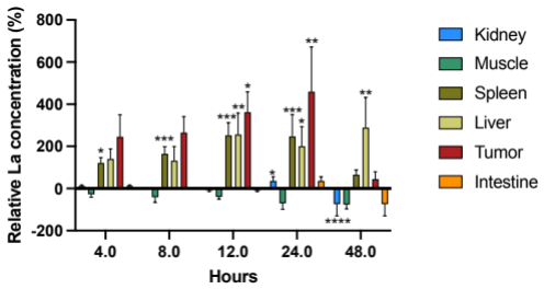
Survives circulation, then collects in tumours through the enhanced permeability and retention (EPR) effect. Peaks in tumour at 24 hours — the window when radiation is delivered.
Surface chemistry drives active uptake into cancer cell lines. Accumulates in phagolysosomes and endoplasmic reticulum, driven by the particle's surface chemistry.
Minimal toxicity across normal and cancer cell lines while the particle is inactive. Only turns on where radiation energy is targeted. No off-target activity.
Mouse biodistribution studies, four to forty-eight hours after injection. Peaks in tumour at 24 hours.
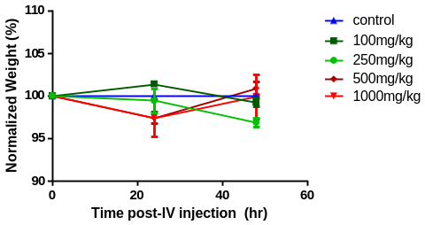
Acute toxicity dose-escalation of the RadioPDT nanoparticle, injected intravenously into C57BL/6 mice.
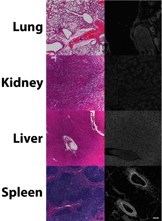
Histopathology across lung, kidney, liver and spleen shows no tissue damage at therapeutic doses.
Normalised weight held flat across the dose escalation. No appreciable toxicity in preclinical models at therapeutic doses.
Post-mortem fluorescence shows clearance through the spleen and liver. Safely broken down by noncancerous cells.
Robust data from in vitro and in vivo models supports the therapeutic potential of the platform. A dual-specific mechanism: the biochemical selectivity of the nanoparticle plus the spatial selectivity of the beam, producing a specific and synergistic kill.
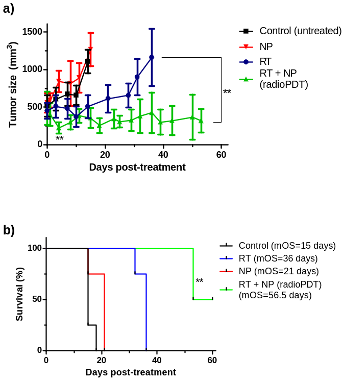
Where radiation alone cannot. Tumour volume held flat while the radiation-only arm keeps climbing. Strong anti-cancer effect beyond radiation alone.
RadioPDT combined with radiation produces significantly greater tumour control than radiation alone. Faster tumour destruction with less cancer resistance.
In preclinical models at therapeutic doses. Only turns on where radiation energy is targeted. No off-target activity.
Preclinical tumour volume and survival data: Control, Nanoparticles alone, Radiation alone, and RadioPDT.
No one else is doing radiation-activated photodynamic therapy in an agent that can actually reach the clinic.
FDA-approved components with proven scalability. PEG-PLGA, LaF3:Ce3+, and PPIX are all well-characterized materials with established safety profiles.
Superior selectivity, and a faster cancer cell kill. Dual specificity: biochemical selectivity of the nanoparticle plus spatial selectivity of the radiation beam.
Fewer permanent radiation "late effects," and it complements the radiotherapy clinics already deliver. No new equipment, no new clinical workflows.